Your one-stop video guide for mastering the Refraction OSCE station
Refraction is a high-yield station in clinical ophthalmology exams such as FRCOphth and Postgraduate Ophthalmology exams. Whether you’re a beginner building your technique or polishing your OSCE performance, this curated video series will guide you step-by-step through the key components of refraction.
🔍 1. Retinoscopy: Mastering the Objective Starting Point
Retinoscopy lays the foundation for accurate subjective refraction. These videos break down technique, neutralization, and how to tackle common challenges.
🎥 How to Perform Retinoscopy – Clear Technique Demo
✅ Tip: Practice observing reflexes at different working distances. Know how to articulate “with” and “against” movements confidently during viva.
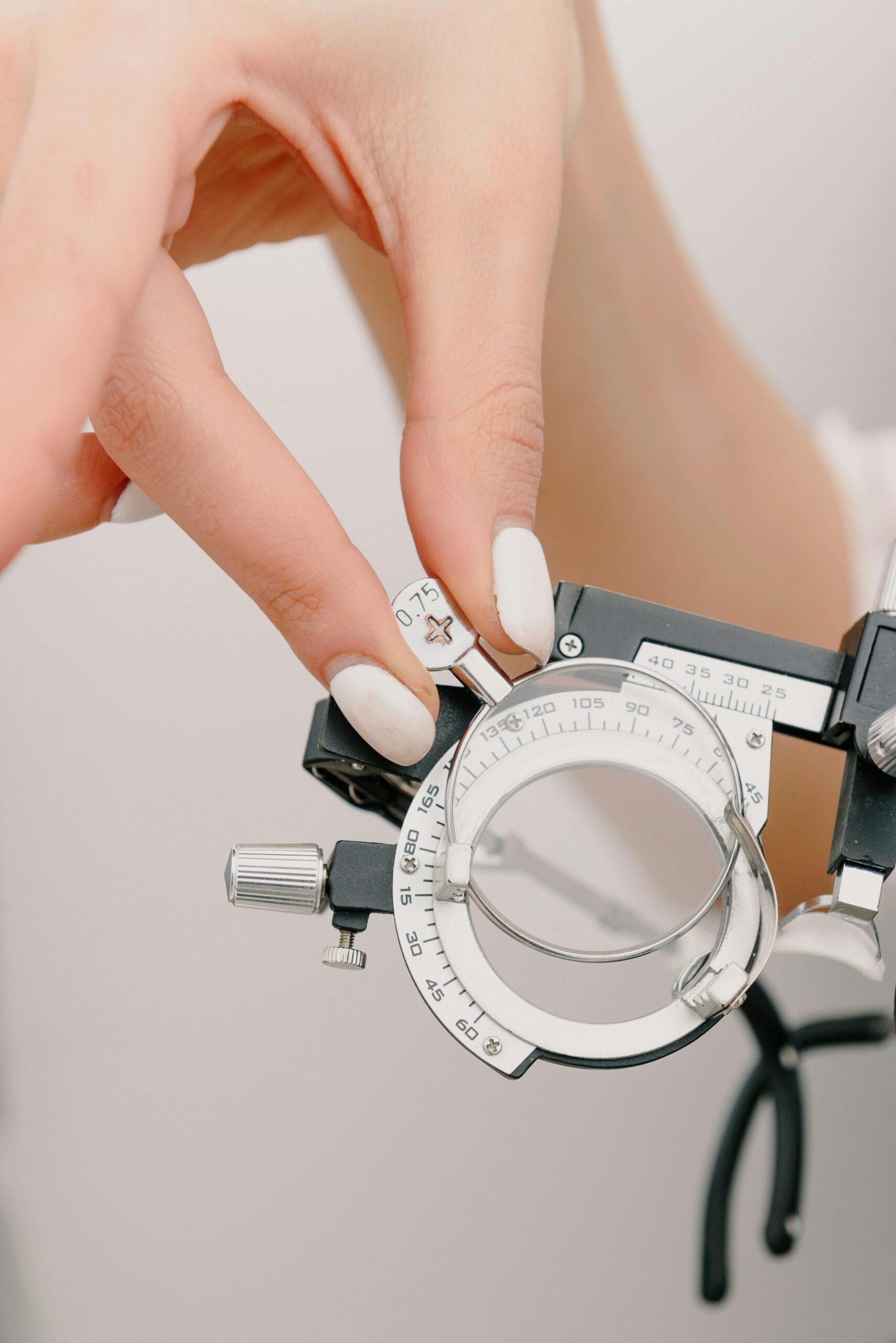
🔵 2. Sphere Refinement: Fine-Tuning Vision
After retinoscopy, spherical power is refined based on patient feedback using bracketing and duochrome tests.
✅ Tip: Know when to stop – over-minusing can reduce clarity and trigger headaches.
🔺 3. Cylinder Refinement: Axis and Power with Confidence
The Jackson Cross Cylinder (JCC) technique is essential to adjust astigmatic correction.
✅ Tip: Always maintain axis before refining cylinder power. Practice communicating each step to simulate exam conditions.
👓 4. Binocular Balancing: Harmonizing the Two Eyes
This step ensures equal accommodation and avoids overcorrection in one eye.
✅ Tip: Master the fogging technique and alternate occlusion methods.
🧾 5. Focimetry: Reading Spectacle Prescriptions Accurately
Learn how to use a manual lensometer (focimeter) – a skill frequently tested in OSCEs and daily clinical life.
- 🎥 Focimetry – Basics
- 🎥 Step-by-Step Lensometer Use
- 🎥 Focimetry OSCE Walkthrough
- 🎥 How to Measure Lens Powers Manually
- 🎥 Interpreting Glasses with Prism
- 🎥 Reading Progressives – Demo
- 🎥 Extra: Focimetry Recap
✅ Tip: Practice identifying optical centers and reading cylindrical components in both plus and minus notation.
💡 Final Tips for the Refraction OSCE
- Always introduce yourself and explain the procedure to the patient.
- Narrate your steps clearly – this scores well during viva or OSCE evaluation.
- Know common scenarios: pseudophakia, high myopia, amblyopia, and anisometropia.
- Practice under time pressure to simulate exam settings.
📌 Bookmark this page and return whenever you need a quick brush-up before an exam or clinic!
✉️ For more posts like this, subscribe or follow @EyeCapsule on social media.